|
|
Parietal Thinning
General Considerations
- Variant seen in parietal bones in which there is thinning of the bones bilaterally
- About 0.4% of normal skulls
- More common in women
- Mean age 72
- Can be progressive
- May be familial
- No emissary vein is transmitted
- Some consider it developmental, some secondary to osteoporosis and others a dysplasia
- Stress produced by chewing has been implicated
Clinical Findings
Imaging Findings
- Oval-shaped lucency seen at upper portion of parietal bone on lateral (sagittal) view
- Loss of diploic space in this region
- Usually bilaterally symmetrical
- Resorption of the outer table
- Inner table is usually intact
Differential Diagnosis
- Parietal foramina

- Posterior portion of parietal bone at upper margin
- If small, they may carry an emissary vein
- When large (several centimeters), they may not carry a vein


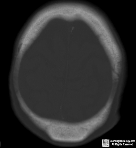
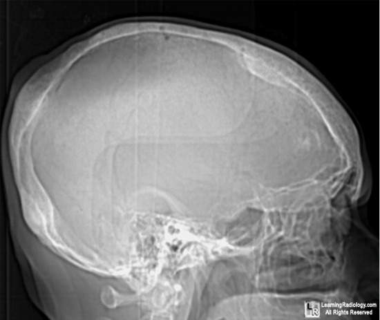
Parietal thinning. Axial CT scan of the skull on the yop shows bilaterally symmetrical thinning of the parietal bones (white arrows). The outer table is resorbed and the diploic space is narrowed. The lateral radiograph on the bottom demonstrates and oval lucency at the vertex of the skull. Again, the outer table is involved while the inner table is intact.
For more information, click on the link if you see this icon
For this same photo without the annotations, click here and here
Diagnostic Neuroradiology Taveras J and Wood E Williams and Wilkins 1964
|
|
|

)